2inch Slackline Kits Aggroline Designed to achieve dynamic movement and bounce at great distances, the AGGRO LINE is ideal for large trees, water lines, tricks or challenging walks. Featuring trampoline-style webbing with rubber grip, Alpha Ratchet with mechanical advantage at long lengths and a backup line system, this kit allows for progression to long lines and advanced tricks. Two-piece Slackline is fully adjustable and easily installed between trees or other sturdy anchor points.Improve balance skills, core strength, and coordination while having fun.Kit includes slackline, Alpha Ratchet, tree protection and safety backup line.Custom-designed trampoline-style webbing is made for slacklining and provides extra bounce for dynamic tricks. Slackline Kits Aggroline,Zen Monkey Slackline,Ourdoor Playing Slackline Training WINNERLIFTING SAFETY EQUIPMENT CO., LTD. , https://www.webestusa.com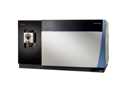 I. INTRODUCTION Cell sorting, microscopic imaging plays an important role in the research of cancer cells, life aging mechanisms and other life science hotspots. The foreign media predicts that under the promotion of cell biology technology, cell discovery services, basic cell biology reagents, flow cytometry,transfection and electroporation, media and serum,microscopy, cell culture equipment, whole cell analysis, and Organization, imaging equipment will produce the largest market scale and technological progress. The flow cytometry and imaging technology have undergone a technological leap over the past period of time. This article presents the new products and technologies introduced in the field of flow cytometry and imaging in the past year to a large number of users.
Flow cytometry is an instrument for the rapid quantitative measurement and analysis of high-speed linear flow cells or biological particles. It mainly includes the flow technology of the sample, the counting and sorting technology of the cells, and the data collection and analysis technology of the computer. The earliest prototype of the flow cytometer was born in 1934. Moldavan proposed the idea of ​​flowing a single suspended red blood cell through a glass capillary, counting it in a bright field with a microscope, and measuring it with a photoelectric recording device. In 1953, Crosland-Taylor designed a flow cell based on the flow of Newtonian fluid in a circular tube. After continuous improvement by Coulter, Parker & Horst, Kamentsky, Gohde, Fulwyler, Herzenberg and others, the photoelectric detection device and cell sorting device were designed to complete the physical connection between the computer and the flow cytometer and the recording of multi-parameter data. The analysis created a cell immunofluorescence staining and detection technology to promote the clinical application of flow cytometry.
In the past 20 years, with the continuous improvement of flow cytometry and its detection technology, new breakthroughs have been made in sample preparation, cell labeling, and software development, making flow cytometry widely used in immunology, biochemistry, and biology. , oncology, hematology and other aspects of research and clinical routine work has become cell sorting, cell cycle analysis, DNA ploidy determination, immunoreactive cell type purification, blood cell typing classification, drug distribution in cells, cell withered Dead and other important research tools in the work.
At present, there are mainly two types of flow cytometers on the market, one is an analytical flow cytometer, and the cell sample is finally analyzed by this instrument and then enters the waste liquid barrel and cannot be recycled. The other is sorting type. Flow cytometry, which can perform both flow analysis and sorting of the target cells for analysis. With the development of technology, multifunctional flow cytometers have emerged, including microscopic imaging flow cytometry. The traditional sorting principle uses charge-based sorting. The new-generation flow cytometer uses fiber optic technology and microchip technology instead of charge-based sorting, so that the cell activity and yield are significantly improved. In recent years, miniaturized personal flow cytometers have emerged. Their main features are low cost, simple operation, low maintenance and use costs, but small-flow instability is the biggest problem.
With the development of cellomics, scientists are eager to explore in-depth and comprehensive information about cells. They provide both group information of cells and individual cell differences. They can not only see the characteristics of cell phenotype, but also can study the Features. The advent of microscopic imaging flow cytometry has revolutionized traditional cell analysis, enabling researchers to simultaneously perform mass cell population analysis and cell image real-time observations.
At the protein level, flow has always been the most commonly used method for single cell analysis. However, because of problems such as cross-coloring, streaming can usually only detect 6 to 10 proteins simultaneously. The emergence of Mass Spectrometry technology has surprised researchers. CyTOF2 mass spectrometry flow cytometry utilizes super-resolution mass spectrometry technology and unique metal-labeled antibodies, which have a qualitative improvement in the number of detection channels and signal quality.
The traditional flow cytometer sheath fluid is very important, the sheath fluid can focus the cells or particles through the laser excitation area. Although sheath fluids have been used in flow mode for decades, fluid dynamics of sheath fluids has increased the complexity of the instrument. The replacement of traditional sheath hydrodynamic focusing systems has become a hot topic of research. The development of fiber optics and microchip technology has made this alternative a reality.
The traditional charge-type flow sorter is characterized by high pressure, high speed, and high charge. It often causes poor cell activity and low yield. Charge-based sorting produces droplets and aerosols, which will produce biosafety for sorting operators. Sexual hazards. The use of microchips for flow cytometry not only ensures the need for high-speed sorting, but also solves the fatal defects of poor activity and low yield of traditional charge-flow sorting cells, and has high biological safety.
As an important analytical instrument for life science research, flow cytometry has always been characterized by high price, high maintenance and use cost, and complicated operation, making it difficult to become a laboratory routine instrument. Recently, miniaturization and personalization has become a hot spot for the development of flow cytometers. Micro-flow cytometers generally have the advantages of low cost, simple operation, low maintenance and low cost of use, and will be widely used in the future.
At present, various microscopic techniques and confocal techniques have been widely used in the field of bioimaging to greatly improve the accuracy of images. The multi-functional integration of cell imaging analysis is the development direction of cell imaging. GE Medical's CytellTM Automated Cell Imaging Analyzer combines the functions of a digital microscope, an image cell analyzer, and a cell counter to simplify the experimental procedure. Bertrand's CytationTM3 cell imaging multi-function detection system integrates an automatic digital microscope and a convenient and flexible microplate reader to meet different testing needs.
The improvement of chemiluminescence imaging technology is mainly due to higher resolution, lower background interference and better sensitivity. In addition, the development of highly sensitive, flexible, and convenient chemiluminescent imaging devices that replace traditional experimental procedures is also a breakthrough point in the future of bioimaging.
II New flow cytometer products launched in 2013 FlowSight Quantitative Imaging Analysis Flow cytometer Mercury Millipore's FlowSight multi-dimensional panoramic flow cytometer launched in October 2013 for the first time from group to individual, from phenotype to function Comprehensive cell analysis has led to a new era of flow cytometry. FlowSight's revolutionary design increases signal-to-noise ratio and the sensitivity of fluorescence detection. With 12 standard detection channels, in addition to the traditional fluorescence intensity information, bright field, dark field, and 10 fluorescent images of “per cell†can be obtained. This instrument has ultra-high detection sensitivity of less than 10 MESF, which has obvious advantages for weak signal detection. FlowSight enables researchers to obtain unprecedented depth of cellular information and expands the application of traditional flow cytometry.
CyTOF2 Mass Spectrometer The CyTOF2 Mass Spectrometer is a new generation of multiparameter flow cytometers from DVS Sciences. It was the first to use metal elements as a marker for flow-through antibodies and dyes, and used mass spectrometry to quantitatively detect labeled cells. On the one hand, the number of flow detection channels has been greatly increased to hundreds, which has increased the amount of information obtained from a single sample; on the other hand, it avoids inter-channel signal interference, greatly simplifies experimental design, and improves data reliability. The increase in the number of signal molecules detected means more accurate observation and categorization of the phenotype within the cell population, a more accurate understanding of the homogeneity of cell lines, and a clearer understanding of intracellular signal transduction networks. At the same time it also provides new research tools for the development of single-cell proteomes.
MACSQuant® Tyto microchip-based flow cytometer MACSQuant® Tyto, introduced by Microtek, is a microchip-based flow cytometry sorter. The traditional charge-type flow sorter has poor cell activity and low yield because of high pressure, high speed, and high charge. With the development of microchip technology, microchip-controlled mechanical valves only need several tens of microseconds to open and close, which has reached the speed of sorting tens of thousands of cells per second by conventional sorters. It not only ensures the need for high-speed sorting, but also solves the fatal flaw of conventional charge-type flow sorting; the sheath-free liquid, closed sample box design avoids the risk of cell contamination, and does not generate droplets and aerosols. Operators have no biosafety hazards.
MoxiFlow Microflow Cytometry MoxiFlow micro flow cytometer introduced by American ORFLO company may be the world's smallest flow cytometer, the size is only 26 × 15 × 15cm, the instrument built-in 4500mAh lithium battery, easy to move in the laboratory and Freely placed; built-in software program, 480 × 320pix color display can clearly show the data results, it uses the Coulter resistance principle and fluorescence detection principle combined detection methods, can be competent for cell activity detection, apoptosis detection, multi-parameter analysis , Cell size detection, fluorescent bead detection, cell cycle detection and other data detection.
HPC-100 Personal Flow Cytometer The HPC-100 Personal Flow Cytometer, introduced by Handyem, Canada, makes fiber optics the first of its kind for flow cytometry systems. The core technology of the HPC-100 is F3 FiberFlow FluidicsTM. Its biggest innovation is the fiber-based microfluidic flow chamber, which enables the same effect as traditional fluid dynamics without the use of consumable-sheath fluids in traditional flow cytometers. This reduces the complexity of the instrument and reduces the cost of the instrument. And eliminate the cumbersome laser spot correction, which has made a great breakthrough in shockproof and portable.
The S3TM Cell Sorter Flow Cell Sorter Bio-Rad Corporation introduced a fully automated flow cytometry sorter, the S3TM Cell Sorter. It contains 1 or 2 lasers and up to 4 fluorescence detectors and 2 scattered light detectors and is detected using standard in-air excitation techniques. Sample sorting can be performed at high speed with accurate droplet breakpoint monitoring and feedback. The S3 cell sorter has a replaceable filter design to optimize the combination of filters and fluorescent dyes. The instrument's small size, built-in flow and temperature control, and the use of unique technology to automate complex sorting setup procedures eliminate the need for user intervention.
III New Imaging Devices Introduced in 2013 CytellTM Fully Automated Cell Imaging Analyzer The CytellTM Fully Automated Cellular Imaging Analyzer released by GE Healthcare's Life Sciences Division combines the functions of a digital microscope, an image cell analyzer, and a cell counter. Its software, BioApps, uses a pre-designed, easy-to-use, automated, modular design that allows cell scientists who have just undergone cytotoxicity testing to rapidly perform routine cell analysis. At the same time, BioApps has great flexibility to meet experienced testers to explore more cell replacement experiments. In combination with the appropriate cell viability kits, data analysis at the single cell level enables the system to have a powerful ability to assess cell viability. This fully automated, multi-functional system covers all steps from imaging to analysis, data visualization, and report generation, greatly simplifying routine experimentation.
CytationTM3 Cell Imaging Versatile Detection System Bertin introduced the CytationTM3 Cell Imaging Versatile Detection System in September 2013. This is a cell imaging multi-functional microplate detection system that incorporates an automated digital microscope in a single system. And convenient and flexible microplate detector. This design provides a large number of cell phenotyping data for plate-based high-density cell research and analysis. Equipped with BioTek's patented microplate detection Hbyrid technology, CytationTM3 also has a filter-based high-sensitivity detection light path and a grating-based highly flexible detection light path to meet the needs of different detection. The scalable fluorescence cell imaging module provides experimenters with rich cell imaging data.
G: BOX Chemi XX9 fluorescence and chemiluminescence imaging system Syngene, UK, has completely upgraded its fluorescence and chemiluminescence product lines and introduced G:BOX Chemi XX9 fluorescence and chemiluminescence imaging systems. The new product has a resolution of 9 million pixels and the system CCD cooling temperature is upgraded to -57°C, which can minimize background interference and produce better imaging results. The CCD chip used has a higher quantum efficiency, which greatly improves the sensitivity of the system detection. G:BOX Chemi XX9 can not only achieve fluorescence, chemiluminescence imaging, but also can be competent for 2D imaging with high resolution requirements. With the new “Edge Light Sourceâ€, 2D glue (including DIGE glue) can be easily analyzed.
C-DiGit digital chemiluminescence scanner In early 2013, LI-COR of the United States introduced the C-DiGit digital chemiluminescence scanner, which provides an efficient and simple technique for Western Blot imaging. The instrument combines the high sensitivity of tablet compression and the convenience of imaging of the dark box CCD imager without the need of darkrooms, films, repeated exposures without multiple times, no need to change the original experimental method, and the sensitivity is equal to the pressure through software control of digital imaging. High quality pictures of tablets. Its large enough aperture makes it possible to efficiently collect light signals. The linear sensor array is a fast scanning imaging system. It is a digital chemiluminescence imaging device that truly replaces the darkroom tablet.